
Vaginal and Uterine Prolapse
What is vaginal or uterine prolapse?
Prolapse happens when the pelvic floor and supporting tissues weaken, allowing the bladder, bowel, vaginal walls or uterus to drop lower than usual. Symptoms can include a bulge, dragging sensation, pelvic pressure, bladder or bowel difficulty, or discomfort with activity.
What treatments can be offered?
- Pelvic floor physiotherapy and lifestyle advice for mild or early symptoms.
- Vaginal pessary fitting to support the pelvic organs without surgery.
- Surgical repair options, including vaginal, laparoscopic or robotic approaches, depending on the type and severity of prolapse.
When to seek advice
Seek advice if you notice a vaginal bulge, persistent pressure, difficulty emptying the bladder or bowel, or symptoms that are limiting walking, exercise or daily comfort.
Interactive Prolapse Animation
Adjust the POP-Q values and treatment option below to view an interactive 3D animation of prolapse severity and surgical repair.



The pelvis is the area of the body between the hip bones. The organs in this area are held in their given location by a set of muscles and other tissues, which make up the pelvic floor. When these muscles and tissues are weak, all the pelvic organs like the uterus, vagina, cervix, bladder and urethra can slip out of place. This is called a prolapse. Almost all of those organs can slip out through the vagina. This is why it is called a vaginal prolapse.
The Vaginal and Uterine Prolapses page answers the following questions:
1. What are vaginal and uterine prolapses?
2. What are the symptoms of a vaginal and uterine prolapse?
3. What are the causes of a vaginal and uterine prolapse?
Vaginal & Uterine Prolapses
1. What are vaginal and uterine prolapses?
Basically, a prolapse is when any organ of the human body drops down out of its correct location. In any of the possible prolapses, that include the vagina and the uterus we can find the following situations:
- Uterine prolapse: in this case, the uterus falls down into the vagina. In the worst scenario, it can come out through the vaginal opening.
- Vagina vault prolapse: the vault of the vagina is the top part of it. It can drop down into the vaginal canal. This occurs most frequently to women who have had a hysterectomy.
- Cystocele: this is when the bladder falls into the vagina.
- Urethrocele: here, the urethra, which is the tube that enables the urine to leave the body, bulges into the vagina. Almost always, this occurs along with a cystocele.
- Rectocele: the rectum, which is the final part of the intestine, bulges entering into the vagina.
- Enterocele: is when the small intestine drops down into the vagina. This prolapse is usually found with a rectocele.
A vaginal prolapse is more common than you may think. It affects 1 in 3 people worldwide.
2. What are the causes of urinary stress incontinence?
In the cases where symptoms present, the patient can feel:
- A feeling of fullness and pain in the pelvic area. This feeling can get worst while standing up.
- Lower back pain.
- Tissues protruding through the vagina.
- Bladder infections and leakage of urine.
- Problems during sex and inserting tampons.
3. What are the causes of a vaginal and uterine prolapse?
In general, pelvic prolapses are caused by the weakening of the pelvic floor muscles. They form a kind of hammock to hold the organs in their place. Those muscles can lose their strength for many reasons. Below are the most common causes of prolapses:
- Pregnancy and vaginal childbirths weaken the pelvic muscles, especially if the woman has experienced a higher-risk pregnancy.
- Some surgeries like hysterectomy or treatments such as radiation can result in prolapses.
- Menopause: in this phase of the life, the ovaries stop the production of oestrogen. This hormone controls the menstrual cycle and keeps the pelvic muscles strong. As the muscles become weaker, women can become more prone to prolapses.
- Extreme physical activity, such as lifting heavy weights or any other activity or condition that increases the pressure in this area.
- Genetic factors can also play a role. Some women have pelvic muscles that are genetically compromised.
4. How are the vaginal and uterine prolapses diagnosed?
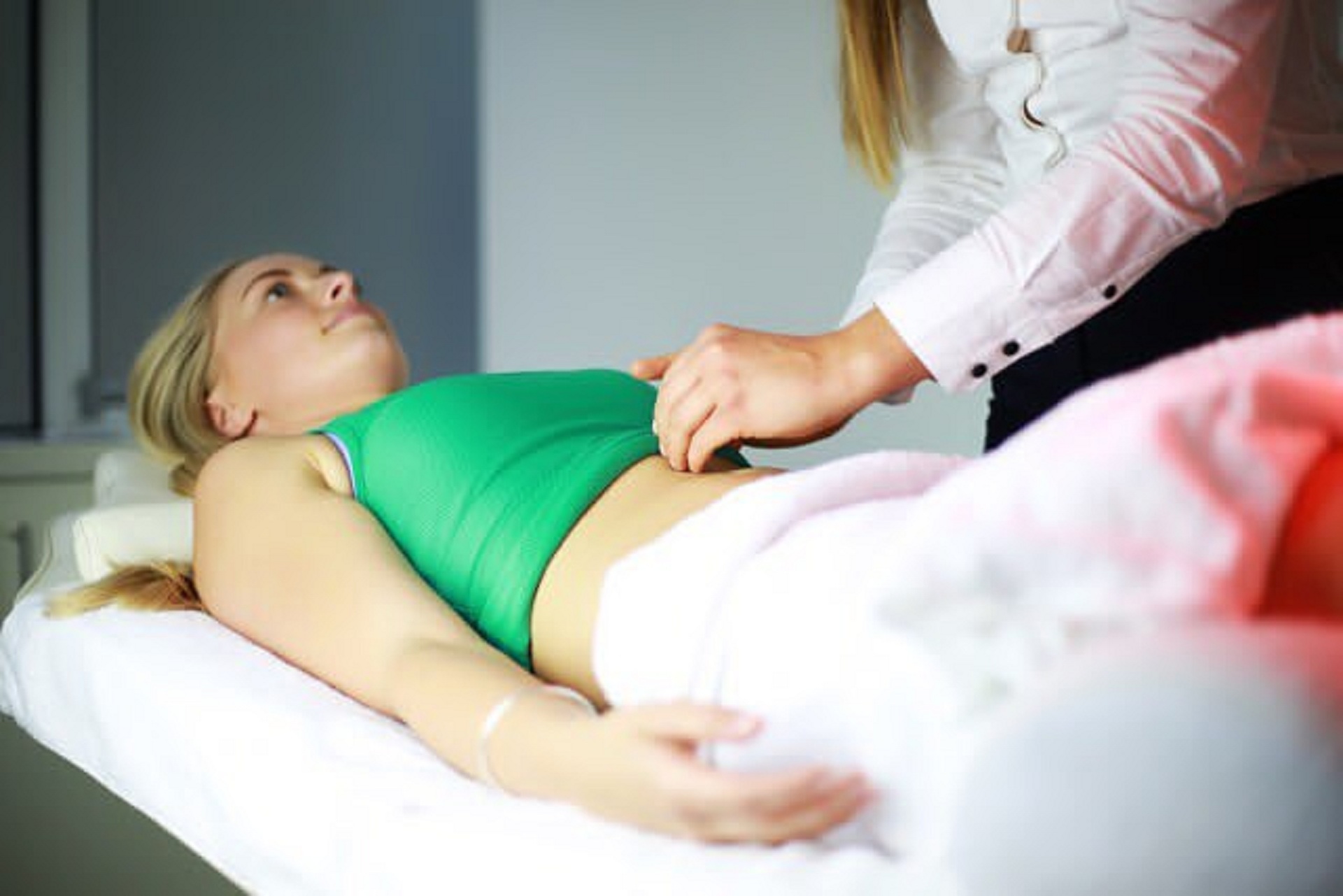
Almost always, a vaginal or uterine prolapse is diagnosed through a pelvic exam. In the exam, the doctor will ask the patient to bear down as if she is doing a bowel movement. The doctor might ask the patient to contract and relax the pelvic muscles used to stop the flow of urine. This simple exam will test the strength of the muscles that support the pelvic organs.
To check the function of the bladder, the doctor can elect to carry out an exam called urodynamic testing. It lasts around 30 minutes and is done using a small catheter to fill the bladder and record the measurements.
Sometimes doctors can carry out a pelvic ultrasound, pelvic floor MRI, or a scan of the abdomen and pelvis to look for problems with the pelvic organs.
5. What are the treatments available for this condition?
The treatment will depend on how weak the pelvic muscles have become. There are non-invasive treatments and surgeries that can resolve the prolapse. At home, women can do Kegel exercises. These will improve the strength of the pelvic muscles. Women just need to tighten pelvic muscles as if trying to stop the flow of urine. These exercises are recommended when women are experiencing little or no symptoms.
A pessary can be used. This is a kind of plastic or rubber ring that will be inserted inside the vagina to support the bulging tissues. It has to be removed regularly for cleaning. Another technique to treat prolapses is administrating oestrogen contained in a cream, suppository ovules or rings into the vagina, to restore the strength of the pelvic muscles.
ProlapLase® is a unique, innovative and non-invasive Er:YAG laser treatment for prolapses. It uses the gentle, non-ablative photo-thermal effects of Fotona’s SMOOTH mode to tighten the tissue and contract the vaginal canal.
ICI 2022 Surgical Pathway for Pelvic Organ Prolapse
This evidence-based pathway supports discussion between patients and their specialist. It is general guidance only; the most suitable treatment depends on the type of prolapse, symptoms, general health, previous surgery, personal priorities and examination findings.
Key pathway findings
- Reconstructive or obliterative surgery: reconstructive surgery aims to restore vaginal support. Obliterative surgery can be effective for older or medically complex patients who do not wish to retain vaginal sexual function.
- Anterior vaginal prolapse: vaginal repair using the patient’s own tissue and sutures is generally the preferred surgical approach.
- Posterior vaginal prolapse (rectocele): native-tissue vaginal repair is generally preferred.
- Apical support: support of the vaginal apex should be considered when anterior or posterior vaginal repair is performed.
- Vault prolapse after hysterectomy: sacral colpopexy is generally preferred, with a vaginal colpopexy representing a reasonable alternative for some patients.
- Uterine prolapse: both hysterectomy and uterine-preserving hysteropexy may be considered. Vaginal hysterectomy combined with apical support is often the preferred option on medical grounds alone.
- Ovaries and fallopian tubes: removal of both tubes and ovaries should be discussed with post-menopausal patients undergoing hysterectomy.
- Stress urinary incontinence: patients with symptomatic or occult stress incontinence should discuss whether continence surgery should be performed at the same time as prolapse surgery.
Adapted from the International Consultation on Incontinence (ICI) 2022 prolapse surgery pathway published by Professor Christopher Maher’s Urogynaecology service.